Plant Cell Viewed Under Light Microscope : Lab Manual Exercise 1 - The part of the cell which surrounds and protects the contents of the cell is called the
byHalina Lynde-
0
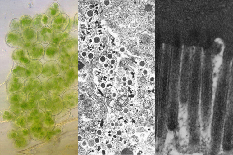
Plant Cell Viewed Under Light Microscope : Lab Manual Exercise 1 - The part of the cell which surrounds and protects the contents of the cell is called the. These cell organelles perform specific functions within the cell. A typical plant cell viewed under a compound light microscope reveals the many different parts that have different functions. Thereof, what does a animal cell look like under a microscope? These cell organelles perform specific functions within the cell. You can see most bacteria and some organelles like mitochondria plus the human egg.
Filed of view is about 1.2mm wide stem of wood dicotyledon, half cross section under microscope. Aims of the experiment to use a light microscope to examine animal or plant cells. The main onion cell structures are quite easy to observe under medium magnification levels when using a light microscope. Some of the cell organelles that can be observed under the light microscope include the cell wall, cell membrane, cytoplasm, nucleus, vacuole and chloroplasts. Read the section cell biology and microscopy on p.3 in your text book.
Cell Organelles Science Learning Hub from static.sciencelearn.org.nz If a plant cell and an animal cell are observed under a microscope, what are the characteristics of the cells that enable you to identify the cell as a plant cells? Xylem cells are dead, elongated, and hollow. The main onion cell structures are quite easy to observe under medium magnification levels when using a light microscope. Aims of the experiment to use a light microscope to examine animal or plant. The xylem is responsible for keeping a plant hydrated by transporting water upward from the roots. Using a light microscope once slides have been prepared, they can be examined under a microscope. Filed of view is about 1.2mm wide stem of wood dicotyledon, half cross section under microscope. On the other hand, eukaryotic cells are more complicated in that they contain a nucleus and many specialized organelles.
The smallest bacteria can't be seen with that magnification.
Cell structures as seen under the light microscope the structures within the cell are referred to as organelles. Some of the cell organelles that can be observed under the light microscope include the cell wall, cell membrane, cytoplasm, nucleus, vacuole and chloroplasts. Beneath a plant cell's cell wall is a cell membrane. See how a generalized structure of an animal cell and plant cell look with labeled diagrams. The cytoplasm contains organelles suspended in fluid. Using a light microscope once slides have been prepared, they can be examined under a microscope. Also know, can rough endoplasmic reticulum be seen. Xylem cells are dead, elongated, and hollow. If a plant cell and an animal cell are observed under a microscope, what are the characteristics of the cells that enable you to identify the cell as a plant cells? (flinn scientific, inc, 2013) the plant cells in this experiment were obtained from the epidermis of an onion. Using a light microscope, one can view cell walls, vacuoles, cytoplasm, chloroplasts, nucleus and cell membrane. Scientists and technicians often use light microscopes to study cells. You can not see the very smallest bacteria, viruses, macromolecules, ribosomes, proteins, and of course atoms.
Microscopy and stained specimens engage students visually as they learn about plant anatomy, a topic covered in many biology and introductory science courses. Light microscope slide with the microsection of a wood stem with vascular bundles. For plant cells, there is a cell wall. Plant material (stained) from a garden water sample, likely sphagnum moss, under the microscope; See the image below illustrating the human cheek cells about 80 µm wide (scale bar is 50 µm).
Section 5 Cells View As Single Page from www.open.edu The major parts of a cell are the nucleus, cytoplasm, and cell membrane. Parts of the light microscope Light plant cell microscope light png clipart free cliparts. Focus your slide on low power. For plant cells, there is a cell wall. Under a microscope, plant cells from the same source will have a uniform size and shape. Animal cell as shown above plant cell as shown above Some plant cell organelles are too.
We offer a wide range of products including foscarini, foscarini and many more.
The part of the cell which surrounds and protects the contents of the cell is called the Similar to the cheek cells, the onion cells need a biological stain to be viewed under the light microscope and for this iodine was used as it binds to the polysaccharides staining the nucleus a brown colour, while leaving the. Under a microscope, plant cells from the same source will have a uniform size and shape. In this activity, students section plant material and prepare specimens to view under a brightfield microscope. Cell is a tiny structure and functional unit of a living organism containing various parts known as organelles. The smallest bacteria can't be seen with that magnification. Identify root hair cells as seen under the light microscope and photomicrograph. Scientists and technicians often use light microscopes to study cells. Plant material (stained) from a garden water sample, likely sphagnum moss, under the microscope; Prokaryotic cells are very simple and lack a nucleus or membrane bound organelles and are small in size. Aims of the experiment to use a light microscope to examine animal or plant. Microscopy and stained specimens engage students visually as they learn about plant anatomy, a topic covered in many biology and introductory science courses. Aims of the experiment to use a light microscope to examine animal or plant cells.
If we cut a longitudinal section of the sunflower stem, we will be able to see the structure of the trachea and tracheid under the high power compound light microscope. You can not see the very smallest bacteria, viruses, macromolecules, ribosomes, proteins, and of course atoms. Prokaryotic cells are very simple and lack a nucleus or membrane bound organelles and are small in size. Under a microscope, plant cells from the same source will have a uniform size and shape. Some plant cell organelles are too.
What Is A Diagram Of A Plant And Animal Cell Under An Electron Microscope Quora from qph.fs.quoracdn.net We offer a wide range of products including foscarini, foscarini and many more. Prokaryotic cells are very simple and lack a nucleus or membrane bound organelles and are small in size. (flinn scientific, inc, 2013) the plant cells in this experiment were obtained from the epidermis of an onion. In this activity, students section plant material and prepare specimens to view under a brightfield microscope. Using a light microscope, one can view cell walls, vacuoles, cytoplasm, chloroplasts, nucleus and cell membrane. Light microscope slide with the microsection of a wood stem with vascular bundles. Microscopy and stained specimens engage students visually as they learn about plant anatomy, a topic covered in many biology and introductory science courses. Onion epidermis with large cells under light microscope clear.
(flinn scientific, inc, 2013) the plant cells in this experiment were obtained from the epidermis of an onion.
You can see yeast cells, animal cells, and plant cells pretty well with a 400x magnification (assuming 10x eyepiece and 40x objective lens). Living organisms are made of cells. Become familiar with the use of the compound light microscope to better understand cell biology. If a plant cell and an animal cell are observed under a microscope, what are the characteristics of the cells that enable you to identify the cell as a plant cells? The major parts of a cell are the nucleus, cytoplasm, and cell membrane. Cell structure hydrilla, view of the leaf surface showing plant cells under the microscope. Thereof, what does a animal cell look like under a microscope? For plant cells, there is a cell wall. You can see most bacteria and some organelles like mitochondria plus the human egg. Scientists and technicians often use light microscopes to study cells. Beneath a plant cell's cell wall is a cell membrane. Identify root hair cells as seen under the light microscope and photomicrograph. Under a microscope, plant cells from the same source will have a uniform size and shape.
Thereof, what does a animal cell look like under a microscope? plant cell microscope view. Beneath a plant cell's cell wall is a cell membrane.